Achilles Tendonitis in Marietta, GA — Targeted Relief & Lasting Recovery
Back-of-heel pain, morning tendon stiffness, and burning tightness make every step difficult. Led by Dr. Neha Delvadia, DPM, Podiatry Group of Georgia provides in-office digital X-rays, hands-on biomechanical gait evaluations, and conservative-first restoration protocols tailored to mid-substance and insertional tendonitis at our Marietta & East Cobb clinic.
What Is Achilles Tendonitis & How Is It Successfully Treated Without Surgery?
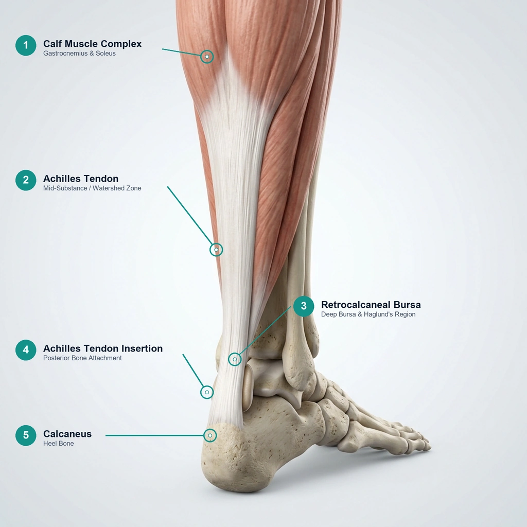
Achilles tendonitis is repetitive micro-strain or degeneration along the thick cord connecting your calf muscles to your heel bone. It presents in two distinct forms: non-insertional (mid-substance) tendinopathy (2–6 cm above the heel) and insertional tendonitis (directly at the bone attachment, often accompanied by bone spurs or Haglund's “pump bumps”). Because chronic tendon pain reflects microscopic collagen wear rather than simple inflammation, passive rest alone rarely cures it. At Podiatry Group of Georgia in Marietta & East Cobb, Dr. Neha Delvadia, DPM prioritizes conservative-first care—using in-office digital X-rays, 3D custom biomechanical orthotics, temporary heel lifts, and flat-ground eccentric rehabilitation to restore tendon fibers before considering surgical intervention.
Why Back-of-Heel Tendon Pain Demands a Targeted Medical Approach
Many patients attempt to treat Achilles stiffness by resting, icing, or following generic calf stretching routines found online—only to find their morning stiffness worsening. When standard home treatments fail, it is usually because the underlying mechanics of the tendon were overlooked:
- The Stretching Trap:Traditional calf stretching off the edge of a step drops the heel into extreme dorsiflexion. For patients with insertional bone spurs or Haglund's bumps, this pinches the inflamed tendon against bone, aggravating micro-tears.
- Tendinitis vs. Tendinosis: Chronic Achilles pain is often not acute swelling (tendinitis), but micro-structural collagen breakdown (tendinosis). Ice and anti-inflammatories temporarily soothe symptoms, but mechanical offloading is required to stimulate tendon fiber repair.
- Rearfoot Pronation & Rotational Shear: Overpronation or flat arches cause the heel bone to tilt inward during gait, creating abnormal twisting shear across the Achilles tendon with every single step that must be neutralized with custom orthotics.
Led by Dr. Neha Delvadia, DPM, Podiatry Group of Georgia takes the guesswork out of Achilles rehabilitation. With in-office digital X-rays and hands-on gait evaluations, we isolate the exact anatomical cause so your treatment begins healing the tendon on day one.
What Type of Back-of-Heel Pain Do You Have?
Because treatments differ fundamentally depending on the anatomical location of your pain, an accurate diagnosis is essential. Compare your symptoms below:
Non-Insertional Tendinopathy
Burning, aching, or localized thickening 1 to 3 inches above the heel bone in the tendon's watershed zone. Often feels stiff in the morning and tender when pinched from side to side.
Insertional Achilles Tendinitis
Pain and tenderness localized right where the Achilles tendon anchors into the posterior calcaneus. Aggravated by firm shoe counters and aggravated if stretched off the edge of a step.
Haglund's Deformity ('Pump Bump')
A visible, prominent bony enlargement on the upper back of the heel bone that rubs painfully against rigid shoe backs, creating redness, friction blisters, or local swelling.
Retrocalcaneal Bursitis
Deep, throbbing ache positioned in the fluid-filled bursa between the front of the Achilles tendon and the top of the heel bone. Often coexists with insertional spurs.
Differential: Plantar Fasciitis
Sharp, stabbing pain concentrated on the bottom or inner curve of the heel during your first morning steps out of bed, rather than the back of the ankle cord.
Differential: Peroneal Tendonitis
Aching or sharp discomfort tracking along the outer edge of the ankle and foot behind the fibula bone, aggravated by lateral cuts, uneven terrain, or unstable shoes.
Urgent Clinical Triage: Did You Feel a Sudden “Pop” or “Snap”?
If you experienced an immediate snapping sensation in the back of your leg during athletic push-off, followed by a visible gap above the heel or the inability to stand up on your tiptoes, you may have suffered an acute Achilles tendon rupture. This requires immediate clinical evaluation. Please call our Marietta office at 404-806-3731 to book your evaluation immediately!
Comprehensive Non-Surgical Solutions for Achilles Tendonitis
Dr. Delvadia utilizes in-office digital imaging and rearfoot biomechanical testing to resolve Achilles pain at its mechanical origin—prioritizing conservative, restorative protocols.
In-Office Digital X-Rays
Immediate weight-bearing views evaluate posterior calcaneal spurring, quantify Haglund's bony prominence, and detect intratendinous calcification on day one.
3D Custom Biomechanical Orthotics
Precision prescription orthotics correct excessive subtalar pronation and rearfoot wobble, neutralizing the rotational shear and twisting forces placed across the tendon with each stride.
Therapeutic Heel Lifts & Offloading
Temporary clinical heel lifts mechanically shorten the gastrocnemius-soleus lever arm, instantly decreasing peak tensile pull across damaged tendon fibers during healing.
Pneumatic CAM Walking Boots
For severe acute flare-ups or suspected partial micro-tears, medical-grade pneumatic boots completely immobilize the ankle mortise while keeping you safely ambulatory.
Flat-Ground Eccentric Rehabilitation
Evidence-based eccentric loading protocols specifically tailored to your diagnosis—performing drops to flat floor level for insertional cases to protect against bone impingement.
Footwear & Heel Counter Modifications
Targeted athletic footwear prescription focusing on optimal heel-to-toe drop ratios (10–12 mm) and unconstricted, soft heel collars that eliminate friction against bony spurs.
When Conservative Care Isn't Enough: Complete Surgical Continuity
While the vast majority of our patients recover completely with conservative offloading and biomechanical support, chronic cases involving prominent calcaneal spurs, recalcitrant Haglund's deformities, or high-grade tendon degeneration may occasionally require surgical evaluation. Because Dr. Neha Delvadia, DPM is a residency-trained podiatric surgeon, you never have to be referred out to an unfamiliar hospital system or start over with a new provider. If surgery is ever needed, the same doctor who knows your foot and athletic goals guides your outpatient surgical care from diagnosis to full recovery.
Your Diagnostic & Treatment Journey
A structured, evidence-based pathway designed to protect your tendon, eliminate mechanical strain, and return you safely to active mobility.
Palpation & Biomechanical Exam
Dr. Delvadia pinpoints the exact site of pathology (mid-substance vs. insertional), measures ankle dorsiflexion, and performs the Thompson squeeze test to verify tendon continuity.
In-Office Digital Imaging
High-resolution digital X-rays evaluate the posterior calcaneus to check for bone spurs, Haglund's deformity, or calcific tendinosis on your very first visit.
Targeted Conservative Protocol
Receive a personalized non-surgical treatment plan—combining 3D custom orthotics, temporary heel lifts, CAM boot offloading if indicated, and flat-ground eccentric exercises.
Gradual Return & Surgical Continuity
Progress safely back to running, tennis, or daily walking. If chronic severe cases fail conservative care, Dr. Delvadia can discuss in-house surgical options without external referrals.